It has been several years since I last looked at pathology slides under a microscope, identifying the different cells and patterns that constitute normally functioning organs and understanding mechanically how structure dictates function. The specialized myocytes of the heart contract in an interwoven manner to orchestrate a contraction. The glandular cells of the thyroid secrete thyroid hormone. The osteoclasts and osteoblasts constantly break down and build up bone in an internal remodeling process. The uterine wall thickens and then sheds with each ovarian cycle in response to hormones. Each cell has its own purpose and works in harmony with the other cells of a specific organ to allow us to live, unaware of the constant machinery within. Pathology is the disruption of the normal structure and thus function of these cells. Diseases can be diagnosed by viewing this distortion under the microscope, in comparison to the norm.


Beyond plain sections of tissue on a slide, there are many ways to observe pathology: fluorescent staining (FISH), staining for various microbial organisms, electron microscopy to delineate finer details, as well as other methods such as radiologic exams (CT scans, PET scans, etc.) These images can be striking for two very different reasons: they can diagnose a very serious disease such as cancer, and they can be aesthetically beautiful.

The macroscopic beauty of human medicine was profound in the Body Worlds exhibit which I first attended in college. One striking piece from the exhibit that I will never forget was the cross-section of a woman and her unborn fetus still within her uterus. She knew she was going to succumb to an incurable illness before giving birth and chose to donate her body to the exhibit so that others could learn. I have also been fortunate to handle the beauty of the human body in medical school during Gross Anatomy, when we dissect cadavers. But the aesthetic beauty of disease is something alluring and unsettling, as one cannot separate the images from the fact that they were obtained from a person inflicted with a disease.

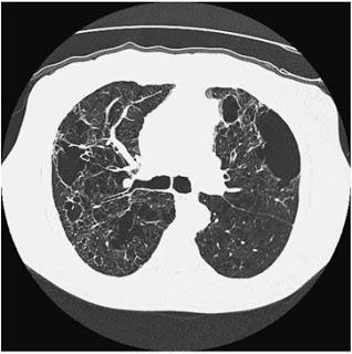
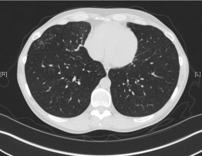
In Hidden Beauty: Exploring the Aesthetics of Medical Science, a large collection of beautiful albeit pathological images are compiled to serve as both art and education. As one of the authors, pathologist Dr. Iacabuzio-Donahue, points out, most people better understand concepts when they are presented visually. A complex disease can be explained by pictures and diagrams. I find that drawing even rough representations for patients allows them to have a clearer understanding of what is going on. Showing them a CT scan of their lungs damaged by COPD is far more poignant than explaining in words what smoking does to the lungs. Likewise, showing people how the normal becomes abnormal as seen under a microscope can be very useful.
As difficult as some of the images may be to experience at first (for example, the cross-section of a fetal hand from an ectopic pregnancy), the book includes explanations of what disease the picture is portraying. If anything, these images remind me of the beauty and symmetry of our bodies. For all the scientific knowledge and advancements of our current day, the basic molecular level of cells has alway struck me as something truly divine.

For a gallery of images from Hidden Beauty, click here.

